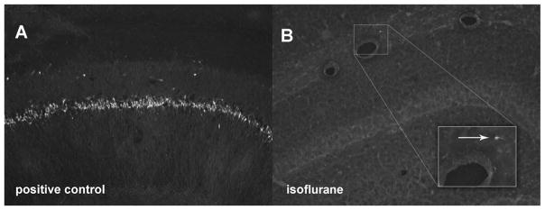
Fig. 3.
FluoroJade (FJ) stain of the hippocampus of 16-month-old rats 16 h after isoflurane (n = 6) or no isoflurane (control, n = 6). Extensive cell death in the CA-1 area of a positive control animal injected with kainic acid (10 mg/kg) 16 h before transcardiac perfusion (A). Almost complete absence of cell death in the hippocampus of a representative isoflurane-treated animal (B). Magnified view of one FJ-positive cell (arrow). Sham-anesthetized animals likewise had no significant detectable cell death (data not shown).

