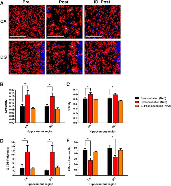
Figure 6.
Incubation of HMW oAβ releases Aβ species that enhance the activation of hippocampal microglia in vivo. The incubated SEC void volume fraction from brain AD1 induced morphological changes in contralateral hippocampal microglia 48 h after injection into lateral ventricle. A, Microglial alterations were analyzed by immunohistochemical morphometry. B, The microglia were analyzed by unbiased automated quantification of the following: B, circularity (4π × [area]/[perimeter]2, with 1.0 indicating a perfect circle); C, solidity ([area]/[convex area], with a maximum value of 1.0); (D) %CD68/microglia; and (E) no. of branches/microglia. Preincubation (black, n = 6), postincubation (red, n = 7), and ID postincubation (orange, n = 2) samples were microinjected into wild-type adult mice. Asterisk (*) indicates statistically significant differences; p values provided in the text.

