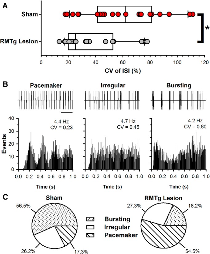
Figure 3.
Effects of RMTg lesions on the basal firing properties of midbrain DA neurons. A, Distribution of ISI CV for neurons from sham (n = 23) and RMTg-lesioned (n = 22) rats superimposed on box-and-whisker plots illustrating the median, IQR, and 5–95% range. CV was significantly reduced in RMTg-lesioned rats. *p < 0.05, Mann–Whitney test. B, Representative samples of spike train event markers associated with pacemaker, irregular, and bursting DA cells (top) with corresponding autocorrelograms (bottom). Note that, despite having similar firing rates, these neurons have different CVs. Scale bar, 1 s. C, Pie charts illustrating the prevalence of pacemaker, irregular, and burst-firing neurons in sham-operated (left) and RMTg-lesioned (right) rats.

