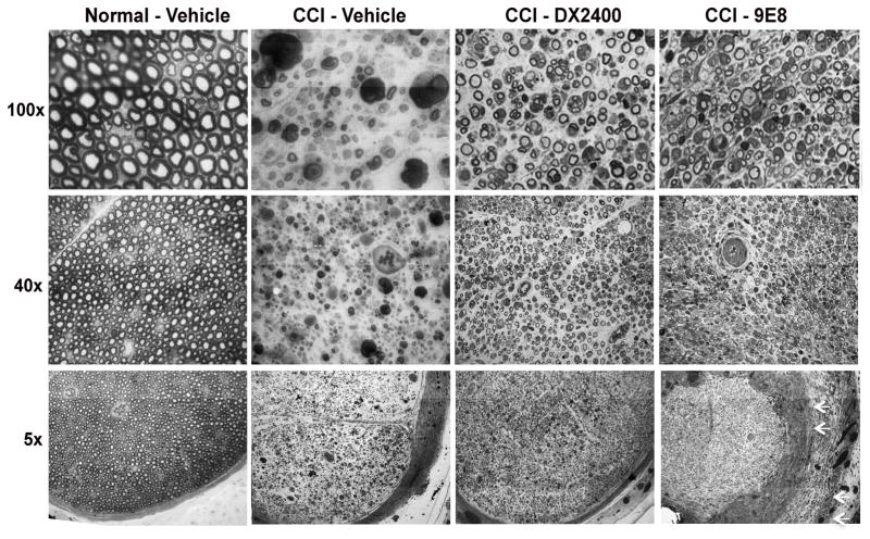
Figure 4. Neuropathology of MT1-MMP inhibition in CCI nerve.
Methylene Blue Azure II staining in 1-μm-thick sciatic nerve sections after i.n. treatment with hAb-DX2400 (1.0 mg/ml), mAb-9E8 (1.0 mg/ml), or PBS (n=4) after completion of von Frey testing in Fig. 3, day 9 post-CCI. Representative micrographs of n=3/group, shown at 5x, 40x and 100x objective magnification. Normal (contralateral) nerve after vehicle injection showed intact nerve morphology. Features of Wallerian degeneration (axon degeneration, edema, myelin ovoids and immune cell infiltration) were observed in CCI nerves after IgG1 treatment. A greater number of uncompromised axons and fewer myelin ovoids were observed in CCI nerves treated with hAb- DX2400 or mAb-9E8. Note the disorganized subperineurial structures after mAb-9E8 treatment (arrows).

