Figure 2.
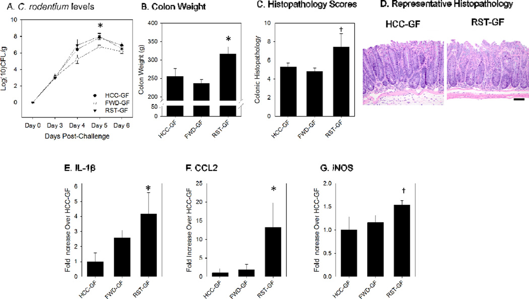
Germ-free mice that received the RST Donor slurry exhibited an elevated inflammatory response to Citrobacterrodentium challenge in comparison with mice that received the microbiota slurry from un-stressed mice. A.) C. rodentium levels were not increased in the RST-GF group over the course of the 6 day study. Colonization levels are displayed in log(10) colony-forming units per gram of fecal pellet. * p<.05 vs. HCC-GF and RST-GF on Day 6 post-challenge. B.) Colon weight was significantly increased at 6 days-post-infection (DPI) in the RST-GF mice. Colon masses are presented in total grams. * p<.05 vs. HCC-GF and RST-GF. C.) Scoring of H&E stained slides for colonic pathology was increased in the RST-GF mice at 6 DPI. † p=.06 (one factor ANOVA). D.) A representative image of the histopathology for an HCC-GF mouse and an RST-GF mouse. E.) Colonic IL-1β mRNA levels were significantly increased in the RST-GF mice over HCC-GF, but not FWD-GF. * p <.05 vs. HCC-GF. F.) Colonic CCL2 mRNA was significantly increased in RST-GF in comparison with both FWD-GF and HCC-GF. * p <.05 vs. HCC-GF and FWD-GF. G.) Colonic iNOS mRNA showed a trend towards a significant increase in the RST-GF mice (p<0.10 in one factor ANOVA). Significance set at p<0.05. All mRNA data are fold increases over HCC-GF using the ∆∆ Ct method and are expressed as the mean +/− standard error.

