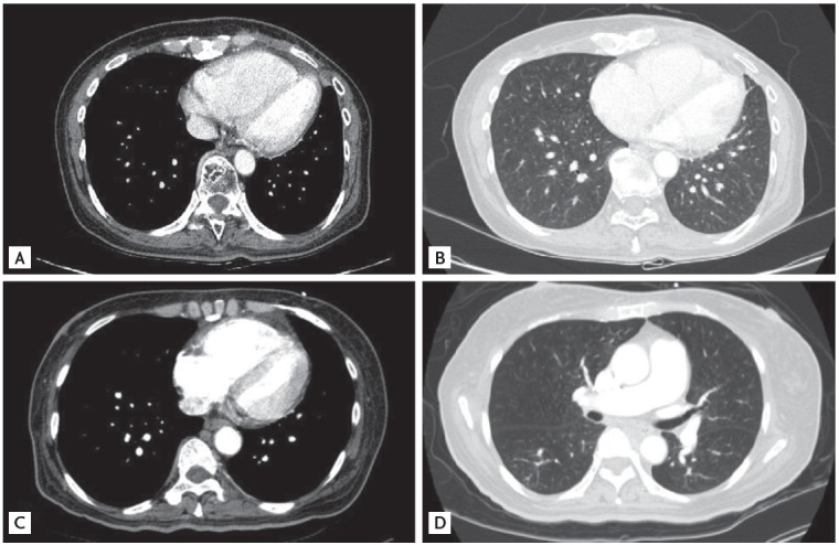
Figure 1.

Computed tomography showed multiple centrilobular nodules with tree-in-bud appearances in cases 1, 2. A mediastinal setting showed a dilated right ventricle and a compressed D-shaped left ventricle in cases 1 and 2, respectively (A, C). A lung setting showed multiple ill-defined centrilobular nodules with tree-in-bud appearances in cases 1 and 2, respectively (B, D).
