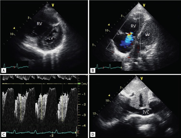
Figure 2.

Echocardiography in case 2. (A) Parasternal short axis view, mid-ventricular plane on transthoracic echocardiography reveals a D-shaped LV throughout the systolic and diastolic period. (B) The 2-dimensional and color Doppler comparative focused image of the apical 4-chamber view showed mild-to-moderate tricuspid regurgitation. (C) The peak tricuspid regurgitation velocity was 3.7 m/sec, pressure gradient 55 mmHg, indicating pulmonary hypertension. (D) The dilated inferior vena cava (IVC) diameter in case 1 was 21 mm without inspiratory collapse. RV, right ventricle; LV, left ventricle.
