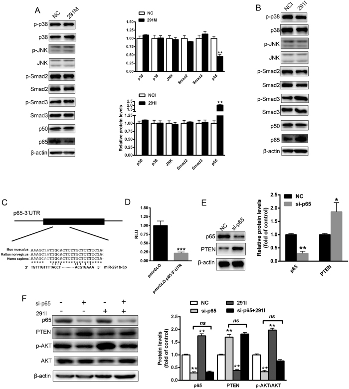
Figure 6. MiR-291b-3p directly targets p65 to upregulate PTEN expression.
(A) The expression or activation of transcription factors and pathways regulating PTEN expression, including p38MAPK, JNK, Smad2/3 and NF-κB (p50 and p65) in the NCTC1469 cells transfected with 291 m or NC. Full-length blots/gels are presented in Supplementary Figure 6 and cropping lines are indicated in red color (left pannel). (B) The expression or activation of p38MAPK, JNK, Smad2/3 and NF-κB (p50 and p65) in the NCTC1469 cells transfected with 291i or NC. Full-length blots/gels are presented in Supplementary Figure 6 and cropping lines are indicated in red color (right pannel). (C) A conserved binding site of the 3′UTR of p65 with miR-291b-3p. (D) Relative luciferase activity in the NCTC1469 cells transfected with reporter constructs containing the 3′UTR of mouse p65 gene. (E) The levels of p65 and PTEN protein in the NCTC1469 cells transfected with a specific siRNA targeting p65. Full-length blots/gels are presented in Supplementary Figure 7A. (F) The levels of p65, PTEN, AKT and AKT phosphorylation in the NCTC1469 cells transfected with p65, miR-291b-3p inhibitor alone or both. Full-length blots/gels are presented in Supplementary Figure 7B and cropping lines are indicated in red color. Data are expressed as mean ± SEM (n = 3 independent experiments). *P < 0.05; **P < 0.01 vs. control group.

