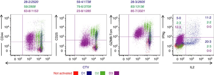
Figure 2.

In vitro differentiation of TCRP1A‐GZMB‐Tom CD8 T cells. CD8 T cells from P1A‐GZMB‐Tom mice labelled with Cell Tracer Violet (CTV) were cultured with 10−7 m P1Ap‐preloaded splenocytes from congenic rag−/− mice (see Materials and methods) for 1 (blue), 2 (green) or 3 (bordeaux) days. Non‐activated CD8 T cells (red) were cultured for 1 day in the absence of P1Ap. CTV and GZMB‐Tom fluorescence as well as staining for CD25 and CD44 expression were measured by FACS (see Materials and methods) on the gated CD8 T cells. Interleukin‐2 (IL‐2) and interferon‐γ (IFN‐γ) production were measured by FACS after reactivation of the CD8 T cells for 4 hr with ionomycin and PMA in the presence of monensin followed by fixation and permeabilization. Numbers indicate % positive cells /(in italic) MFI of the positive cells. Results are representative of three and two experiments, respectively, for surface markers and for cytokines.
