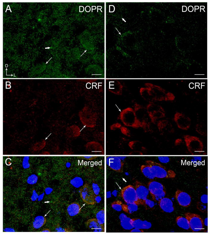
Figure 3.
High magnification confocal immunofluorescence photomicrographs showing labeling for DOPR and CRF in a coronal section through the BLA (A–C) and CeA (D–F). Nuclei were stained by 4′,6-diamidino-2-phenylindole (DAPI). DOPR immunoreactivity was labeled with fluorescein isothiocyanate (green), CRF was labeled with rhodamine isothicyanate (red) and DAPI in blue. A–C. Thin arrows depict labeling for DOPR (A) and CRF (B) with DAPI while thick arrows depict singly labeled DOPR and CRF, respectively, in the BLA. D–E. Thin arrows depict labeling for DOPR (D) and CRF (E) with DAPI in the CeA while thick arrows depict singly labeled DOPR and CRF. Panels C and F show merged image of DOPR, CRF and DAPI. Corner arrows indicate dorsal (D) and lateral (L) orientation. Scale bar, 25 μm.

