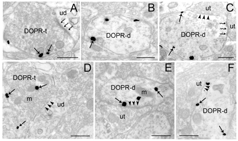
Figure 4.
Ultrastructural evidence for DOPR immunoreactivity in axon terminals and dendrites in the BLA and CeA. Electron photomicrographs showing immunogold-silver labeling for DOPR in the BLA (A–C) and CeA (D–F). A. Immunogold-silver labeling is present in a BLA axon terminal (DOPR-t) which forms an asymmetric synapse (zigzag arrows) with an unlabeled dendrite (ud). Arrows point to DOPR. B–C. DOPR-labeled dendrites (DOPR-d) in the BLA. Panel C shows a DOPR-d that receives an asymmetric (zigzag arrows) and symmetric synapses (arrowheads) from unlabeled terminals (ut). D. DOPR-immunogold-silver labeling can be seen in CeA axon terminal (DOPR-t) that forms a symmetric synapse (arrowheads) with unlabeled dendrite (ud). E–F. DOPR-containing dendrites in the CeA. Both DOPR-d receive a symmetric synapse (arrowheads) from an unlabeled terminal (ut). Arrows depict DOPR-immunogold silver particles. m, mitochondria. Scale bars, 0.50μm.

