FIG 7.
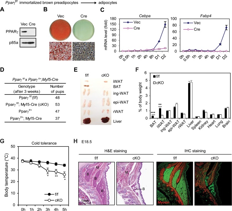
PPARγ is essential for adipogenesis and BAT development. (A to C) Pparγf/f immortalized brown preadipocytes were infected with MSCVhygro expressing Cre. (A) Western blot analysis of PPARγ. p85a was used as a loading control. (B) Seven days after induction of adipogenesis, cells were stained with Oil Red O. (C) Time course qRT-PCR analysis of adipogenic markers Cebpα and Fabp4. (D to G) Characterization of 6-week-old Pparγf/f; Myf5-Cre mice. (D) Genotyping results. (E) Representative pictures of isolated adipose tissues and livers. (F) The relative weight of each tissue and organ is shown as a percentage of body weight. (G) Pparγf/f; Myf5-Cre mice are cold intolerant. Six-week-old mice were housed at 4°C, and their body temperatures were measured every hour (n = 6). *, P < 0.05; **, P < 0.01, ***, P < 0.001. (H) E18.5 embryos were sagittally sectioned along the midline. The sections of the interscapular area were stained with H&E (left panels) or with antibodies recognizing BAT marker UCP1 (green) and skeletal muscle marker myosin (red) (right panels). IHC, immunohistochemistry.

