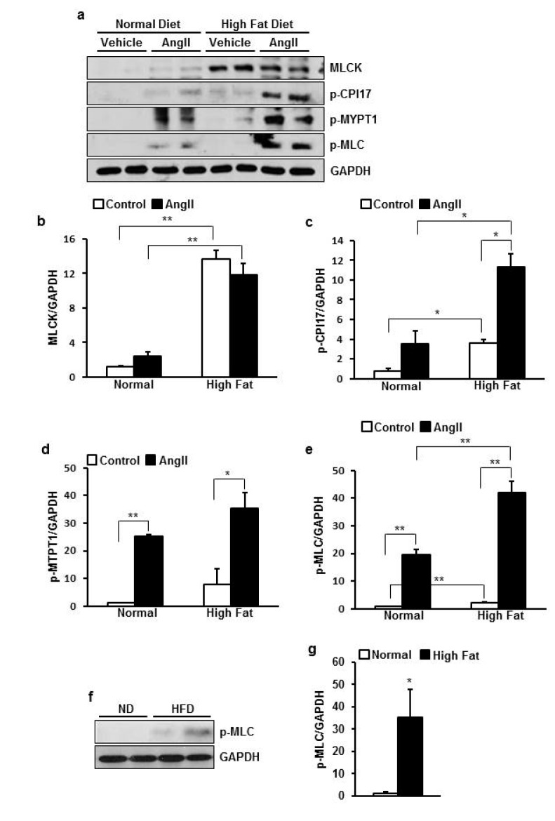
Fig. 5. HFD increased basal expression of MLCK and phosphorylation of CPI-17.
(a) Representative picture of western blot for phosphorylated MLC at Thr18 and Ser19, phosphorylated MYPT1 at Thr853, phosphorylated CPI-17 at Thr38, MLCK, and internal control GAPDH in ND or HFD-fed rat aorta with/without stimulation. (b~e) Summary of protein expression of MLCK (b), p-CPI-17 (c), p-MYPT1 (d), and p-MLC (e). (f) Representative picture of western blot for phodphorylated MLC at resting state in ND and HFD-fed rat aorta (g). Summary of phosphorylation of MLC. Basal expression of MLCK, p-CPI-17, p-MLC was increased by HFD. Ang II-induced phosphorylation of CPI-17 and MLC was also enhanced by HFD. Data are presented as means±SE (n=4). *p<0.05, **p<0.01.

