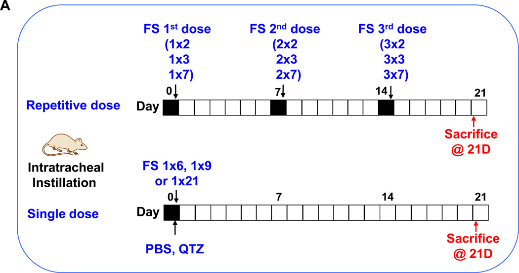
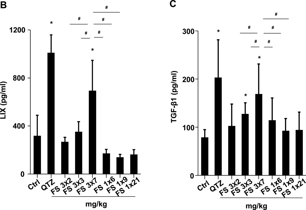
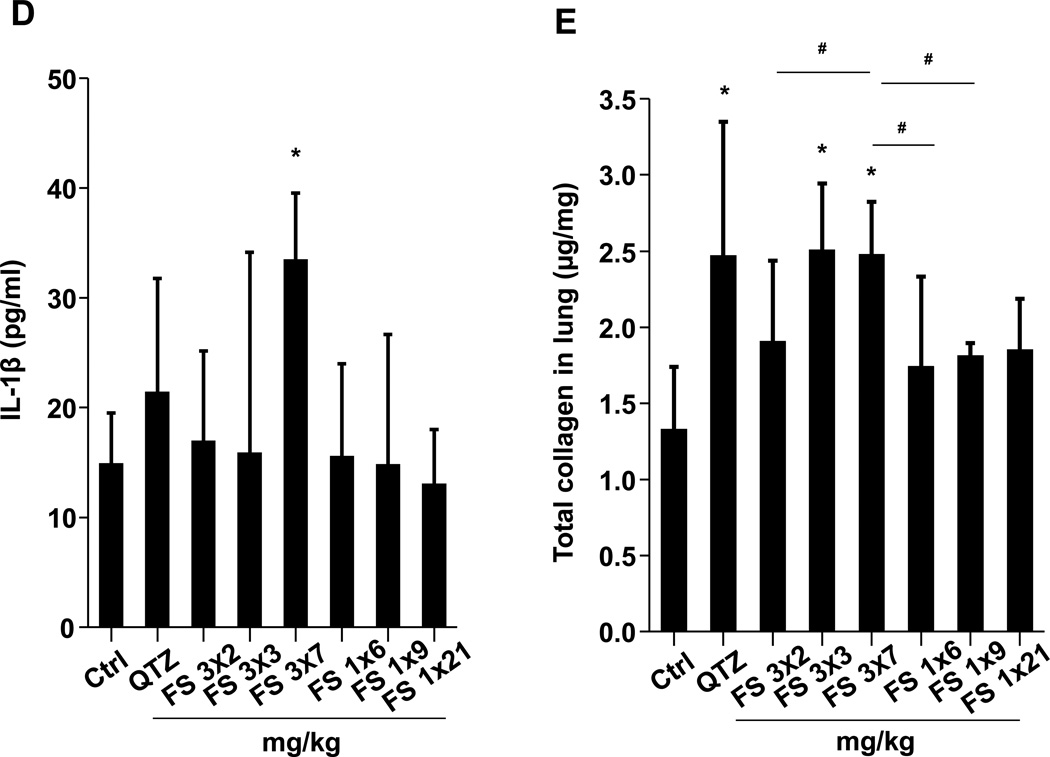
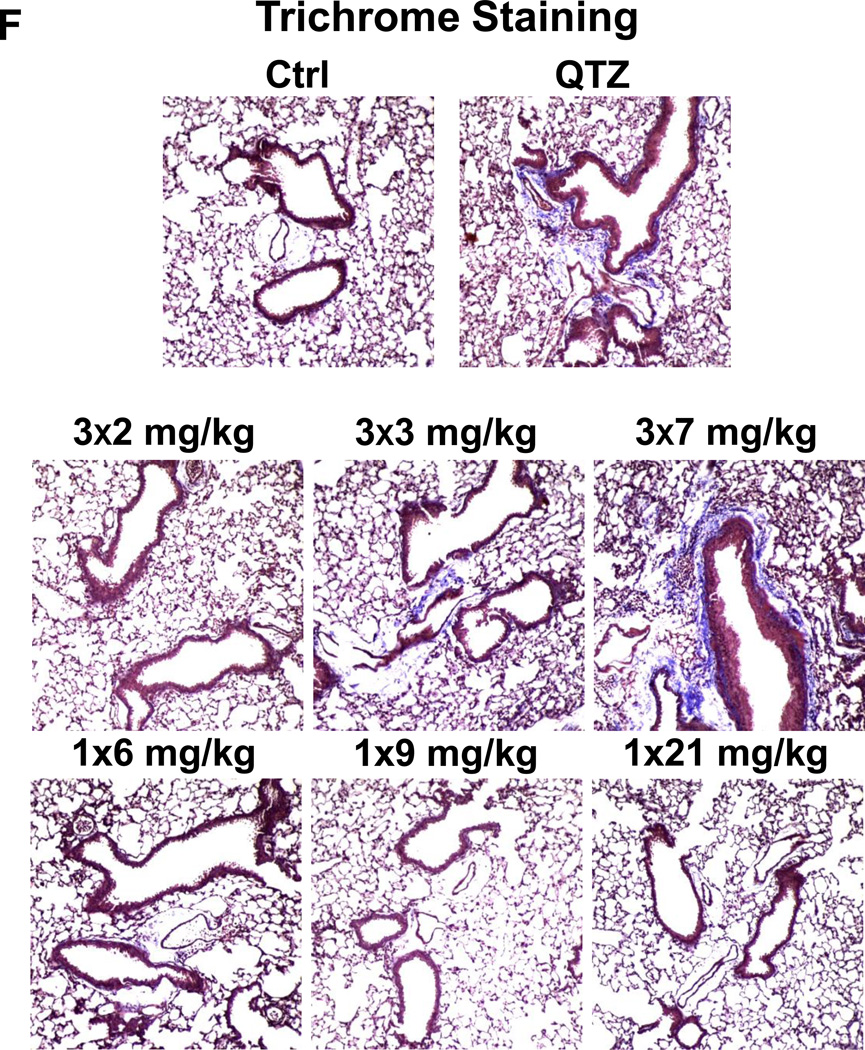
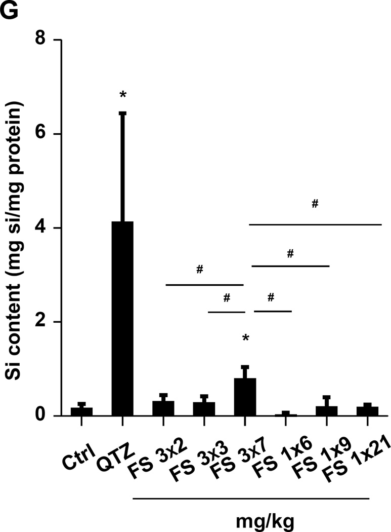
Figure 2. Assessment of sub-chronic pulmonary effects of fumed silica by single vs. repetitive exposures in murine lung.
(A) Schematic presentation of the repetitive exposure conditions in the murine lung. C57BL/6 (n=6) mice were exposed to either 3 doses of 2, 3, and 7 mg/kg, one week apart (i.e., 3×2, 3×3, and 3×7 mg/kg) compared to single dose of 6, 9, and 21 mg/kg by oropharyngeal aspiration. (B-D) On day 21, BAL fluid was collected to determine (B) LIX, (C) TGF-β1, and (D) IL-1β levels. MIN-U-SIL, a natural form of α-quartz (QTZ), was used as a positive control. (E) Total collagen content in murine lung was determined by the Sircol collagen assay. (F) Masson’s Trichrome staining was used to visualize collagen deposition in the lungs of animals treated with fumed silica. Collagen is stained blue in the micrographs. (G) ICP-OES analysis showing silicon abundance in the lung. *p<0.05 compared to control mice. #p<0.05 compared to mice treated with 3 doses of 7 mg/kg fumed silica.

