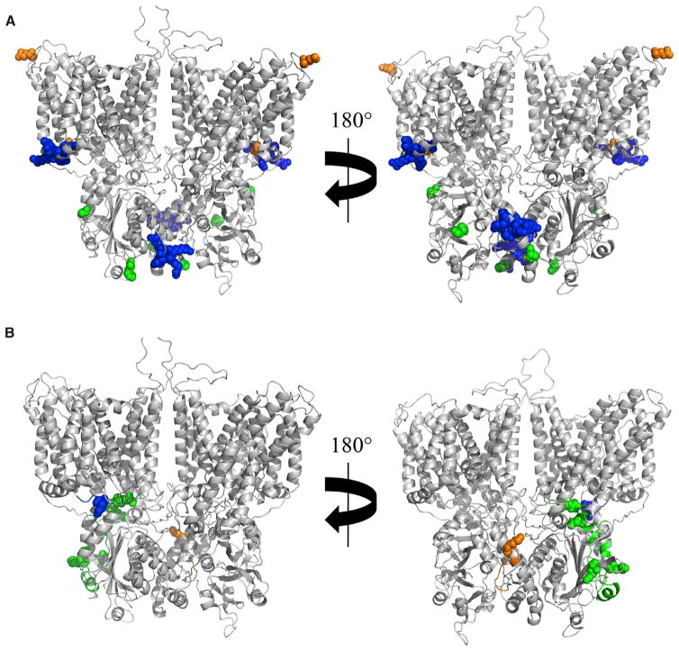
Figure 6. CX-MS of Intact Membranes Suggests Protein-Protein Interfaces Are More Extensive than Defined Previously.
(A) Previously described interaction sites on an AE1 dimer for glycophorin A (residues 658 and 701, orange spheres), protein 4.1 (residues 343–347 and 386–390, blue spheres), and protein 4.2 (residues 40, 130, and 327, green spheres) are highlighted.
(B) New interaction sites on AE1 for glycophorin, proteins 4.1 and 4.2, are shown using the same color coding as in (A).

