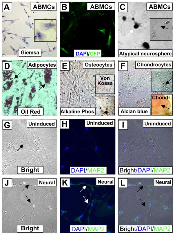
Figure 2.
Canine ABMC tripotency and neural induction. (A) Canine adherent BM cells isolated after 72 h (ABMCs) and stained with Giemsa. Inset shows a higher-power image of the cells. (B) Canine ABMCs transfected with GFP with ~95% efficiency. All cells in this field are positive for green fluorescent protein (GFP). (C) Neural induction of ABMCs after 1 week showing atypical neurospheres (white arrow) and extended dendrites (arrow in inset). (D) ABMCs induced for adipocytes and stained with oil red. (E) Osteocyte differentiation with Von Kossa staining (upper inset) and alkaline phosphatase staining (lower inset). (F) Alcian blue staining of ABMCs induced for chondrocytes in either tissue culture plate (upper inset) or as chondroitin sulfate aggregates (arrow) in a tube 3D culture (lower insert). (G–L) Bright and GFP images of uninduced control ABMCs (G–I) and ABMCs induced for neural differentiation and stained for the neuronal dendrite-specific marker microtubule-associated protein 2 (MAP2). Notice that uninduced cells are negative for MAP2 (H). After 12 days, induced cells demonstrate extended dendrites connecting neural-like cells that are positive for MAP2 (J–L). Nuclei were counterstained using 4′,6-diamidino-2-phenylindole (DAPI). Scale bars: 20 μm (A–L).

