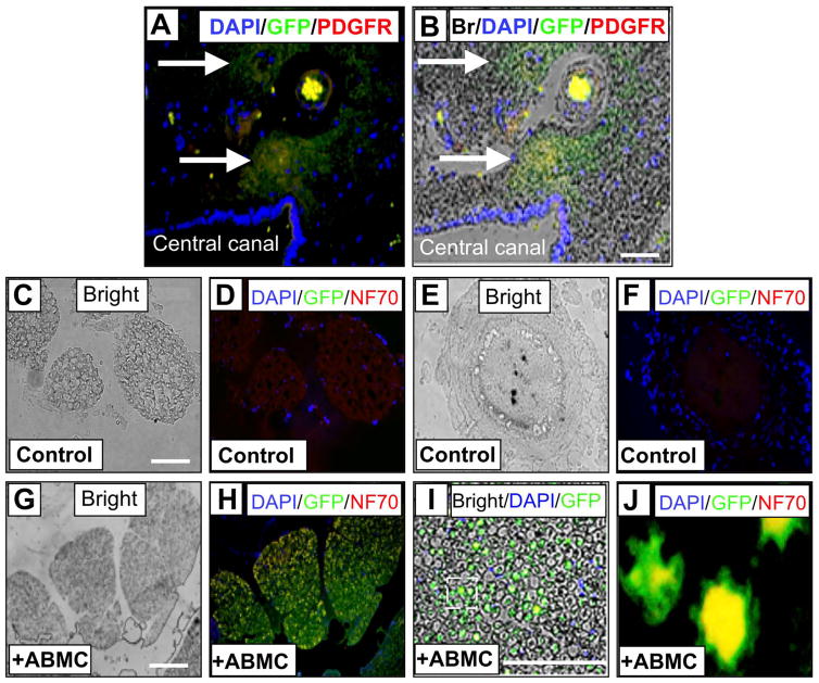
Figure 6.
Immunofluorescence staining of ABMC-derived cells positive for GFP and PDGFR at central canal and white matter spinal cord sections. (A) Fluorescent images of nuclear marker DAPI (blue), GFP (green) as a marker for transplanted canine ABMCs, and PDGFR (red) marker of neural progenitor cells near the central canal and associated with small vessels within the spinal cord. DAPI-positive nuclei lining the central canal are seen at the lower left area. (B) Overlay of the fluorescent images on the bright field showing GFP-positive cells coexpressing PDGFR within the spinal cord gray matter surrounding the central canal. (C–F) Spinal cord section from a control dog. (G–J) Section of spinal cord from dog treated with autologous ABMCs. (C) Bright field section and (D) fluorescent images of nuclear marker DAPI (blue), GFP (green), and NF70 (red) neuronal marker. (E) and (F) Higher magnification images from spinal cord sections of a control dog. No GFP expression was detected, while mild NF70 expression is seen in control sections. (G) Bright field and (H) fluorescent images of white matter in (E) stained with DAPI, GFP, and NF70. (I) Higher-power image of overlay of fluorescent images in (H) on the bright field in (G). (J) Higher magnification of outlined area in (I) showing GFP+ axons with colo-calized NF70 expression (yellow). Scale bars: 50 μm (A–B) and 100 μm (C–J).

