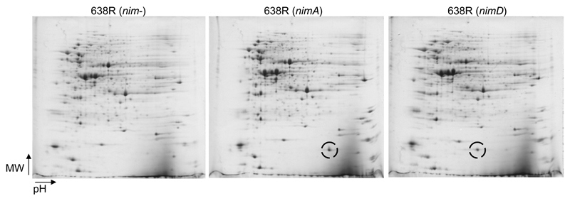
Figure 1.
Representative images of 2D-gels (pH range 3-10 non-linear, 12.5% PAA) from strain 638R, either without nim gene (left image), with plasmid pIP417 (nimA, IS1186), nimA-positive, (middle), or with plasmid pIP421 (nimD, IS1169), nimD-positive, (right). The Nim proteins (encircled) are easily discernible as prominent spots in the lower molecular mass range of the gel. The theoretical molecular weights of NimA and NimD amount to 20.2 kDa and 18.5 kDa, respectively. Directions of increasing molecular mass (MW) and increasing pH are indicated by arrows.

