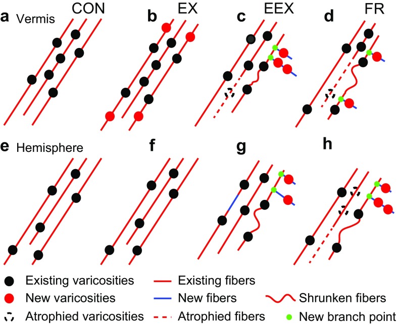
Fig. 9.
A neural model of exercise-evoked NA plasticity in the cerebellar vermis and hemisphere that is consistent with the collected data. a–d Vermis NA fibers and varicosities. e–h Hemisphere NA fibers and varicosities. a, e Intervals between varicosities for CONs are slightly shorter in cerebellar vermis, compared to cerebellar hemisphere (P < 0.01, Student’s t test), but both regions still exhibit similar levels of clustering (P > 0.05, Student’s t test). b, f Mild exercise-evoked addition of NA varicosities to existing fibers in the vermis of EX rats, but not in the hemisphere of EX rats. c, g Relative to CON rats, food restriction-evoked excessive exercise leads to shorter inter-varicosity intervals in both the vermis and the hemisphere of EEX rats. There is a higher frequency of shorter varicosity containing fragments especially in vermis of EEX rats, corroborating the increased clustering among the NA varicosity population. c, g and d, h Food restriction, with or without exercise, increases the proportion of short NA axon fragments in both the vermis and hemisphere. This change could be due to sprouting of new fibers (blue) associated with the emergence of new branch points (green filled circles) that are not captured in the sampled volume, atrophy of existing axons (dotted red lines) or shrunken fibers (wriggly lines). Red circles represent newly added varicosities. Dotted circles represent varicosities lost

