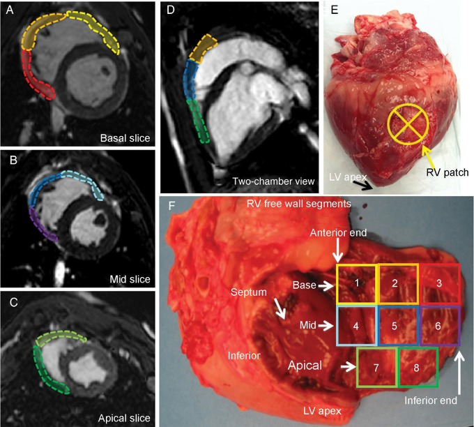
Figure 2:
The right ventricular free wall segments. The right ventricular free wall was divided into eight sections using left ventricle segmentation as a reference: (A) The basal segment and (B) mid segment are further divided into anterior, lateral and inferior segments. (C) The apical slice is smaller compared with other two longitudinal segments and is divided into anterior and inferior segments. (E) An example of macro inspection of the right ventricle patch on the mid-lateral segment. (F) A photograph of the extracted heart with the right ventricle free wall cut at the basal-inferior-apical end and opened. The colour of the eight segments corresponds to A–D. In this specimen, the patch is located in 5, 7 and 8 (i.e. mid apical–lateral wall). Inferior: right ventricular inferior wall; LV: left ventricle; RV: right ventricle; septum: interventricular septum.

