Figure 6:
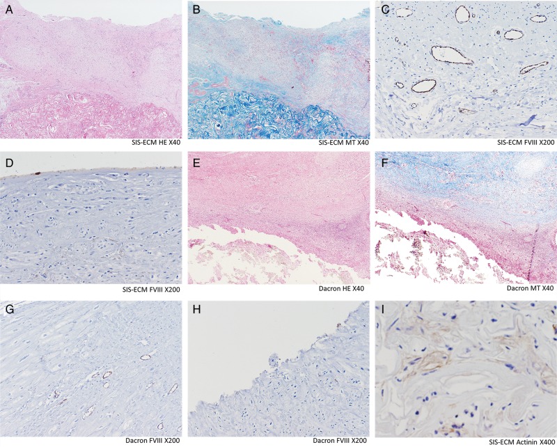
Histology and immunohistochemistry. SIS-ECM (A–D and I): Well-organized cells (shown in A and B) including monolayer of endothelial cells on the endocardial surface (shown in D) and layers of scattered α-sarcomeric actinin-positive cells (shown in I), were observed. The repopulated cells were predominantly observed on the SIS-ECM and extended towards the endocardial surface. Dacron (E–H): Significant infiltration of lymphoid cells and fibrosis is observed (shown in E and F). No endothelial cells were observed in the endocardial surface (shown in G). Capillary density (C and G): The factor-VIII-related antigen-positive capillaries were counted under the ×200 microscopy for capillary density analysis. Vasculogenesis/angiogenesis was significantly more prevalent in SIS-ECM patch. Dacron: Dacron patch; FVIII: immunohistochemistry staining for factor-VIII-related antigen; HE: haematoxylin–eosin staining; MT: Masson trichrome staining; SIS-ECM: extracellular matrix patch derived from small intestine submucosa.

