. 2016 Dec 10;5:e20533. doi: 10.7554/eLife.20533

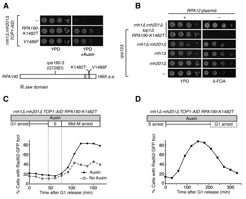
Figure 7. RPA190 mutants allow for repair of R-loop induced damage.

(A) Top: Rpa190-K1482T and -V1486F suppress auxin sensitivity of rnh1∆ rnh201∆ TOP1-AID cells. 10-fold serial dilutions of saturated cultures were plated onto YPD or YPD with auxin. Bottom: Schematic of Rpa190 showing the location of mutations and the jaw domain, as previously published (Engel et al., 2013; Fernández-Tornero et al., 2013). (B) Rpa12 is required in rnh1∆ rnh201∆ TOP1-AID RPA190-K1482T. Cells carrying a plasmid expressing RPA12 and URA3 were plated onto media lacking uracil (-URA, selects for plasmid) or media containing 5-floroorotic acid (5-FOA, selects for plasmid loss). 10-fold serial dilutions are shown. (C) Rpa190-K1482T does not change accumulation of Rad52-GFP foci. Experiment in Figure 4A was repeated on rnh1∆ rnh201∆ TOP1-AID RPA190-K1482T cells. (D) Rpa190-K1482T allows for repair of Rad52-GFP foci. Experiment in Figure 2B was repeated on rnh1∆ rnh201∆ TOP1-AID RPA190-K1482T cells in the presence of auxin.

DOI: http://dx.doi.org/10.7554/eLife.20533.015

Figure 7.

Figure 7—figure supplement 1. Flow cytometry on rnh1∆ rnh201∆ TOP1-AID RPA190-K1482T cells.

Figure 7—figure supplement 1.

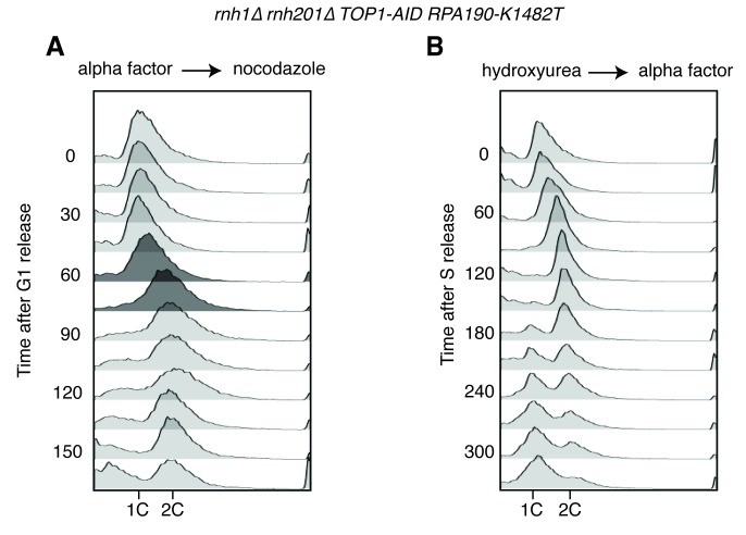
All cells have been treated with auxin for two hours before release and auxin is maintained in the culture after release. (A) Cells are released from alpha factor into nocodazole. Flow cytometry profiles correspond to Figure 6C. (B) Cells are released from hydroxyurea into alpha factor. Corresponds to Figure 6D.
Figure 7—figure supplement 2. Pulsed-field gel electrophoresis of R-loop mutants.

Figure 7—figure supplement 2.

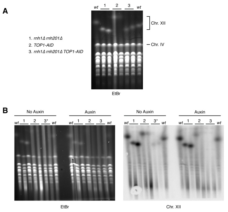
(A) Two independent isolates each of wild-type (wt), rnh1∆ rnh201∆ (1), TOP1-AID (2), and rnh1∆ rnh201∆ TOP1-AID (3) were grown to saturation in YPD and subjected to PFGE. Gel is stained with ethidium bromide (EtBr). (B) The same cells were diluted, allowed to grow to mid-log phase, and treated with or without auxin. Asterisk indicates rnh1∆ rnh201∆ TOP1-AID cells arrested in nocodazole (3*) to ensure cells are in the same part of the cell cycle as those treated with auxin (G2-M arrested). Left: Gel is stained with ethidium bromide (EtBr). Note that replicating chromosomes from dividing cells run poorly and result in smearing – compare to condensed chromosomes from saturated cultures in (A). Right: Southern blot probing the 35S gene in the rDNA locus on chromosome XII. Note the signal in TOP1-AID (2) and rnh1∆ rnh201∆ TOP1-AID (3) is diminished when cells are treated with Auxin.
Figure 7—figure supplement 3. Structural analysis of Rpa190 in the context of the RNA pol I complex.

Figure 7—figure supplement 3.

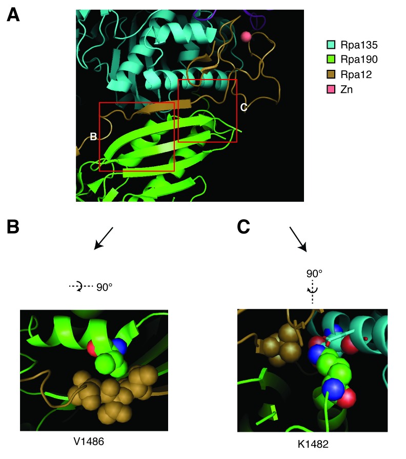
Structure shown is from Protein Data Bank accession number 4C3I, as originally published in Fernández-Tornero, et al. (2013). (A) The ‘jaw’ domain of Rpa190 (green) is shown with the N-terminal zinc-ribbon of Rpa12 (brown) and the ‘lobe’ domain of Rpa135 (cyan). (B) The highlighted region rotated to see residue V1486 in its position on the alpha helix of RPA190 behind the beta sheet in the foreground of (A). Residue V1486 on Rpa190 is shown in spherical space along with residues T49, T50, and T51 of Rpa12. Potential steric clashes arise between these residues when Rpa190-V1486 is modified to phenylalanine. (C) The highlighted region rotated to see residue K1482 behind the beta sheet in the foreground of (A). Residue K1482 of Rpa190 is shown in spherical space along with Rpa135-D304 and Rpa12-V47 in the background. Also in close proximity are Rpa135-E307 and Rpa12-S6 shown as stick models in the foreground. Modifying Rpa190-K1482 to threonine increases the distance between these residues and possibly interrupts acid-base interactions anchored by the lysine residue.