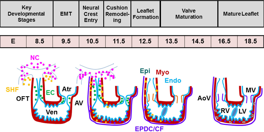
Figure 1. Diagrammatic representation of heart development.
Endocardium (EC) (aqua blue line) forms the endocardial cushions (green circle) via cushion EMT (E9.5). Neural crest (NC) (pink circle) cells migrate into the OFT during E10.5-12.5. Valve leaflets undergoing differentiation (orange color) and maturation (purple color) (E13.5-18.5) are clearly indicated. Only one semilunar valve is shown. SV, semilunar valves; AV, atrioventricular canal; RV, right ventricle; LV, left ventricle

