Fig. 2.

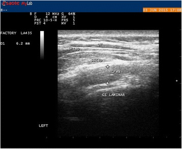
Ultrasound image of the Rcpm at rest in transverse view at C1. The caliper is placed around the midpoint above the C1 lamina betwen the inner and outer borders of the muscle where it was considered thickest by the raters

Ultrasound image of the Rcpm at rest in transverse view at C1. The caliper is placed around the midpoint above the C1 lamina betwen the inner and outer borders of the muscle where it was considered thickest by the raters