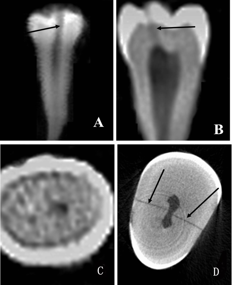
Fig 3. Group C, where both CBCT and PR can detect the crack.
An image of the crack in a premolar tooth is shown. The crack was detected by PR (A) (arrow). The crack was detected by CBCT in the sagittal plane (B) (arrow). No crack was detected by CBCT in the same sample in the horizontal plane (C). The crack was detected by Micro-CT in the horizontal plane (D) (arrow).

