
Figure 4.
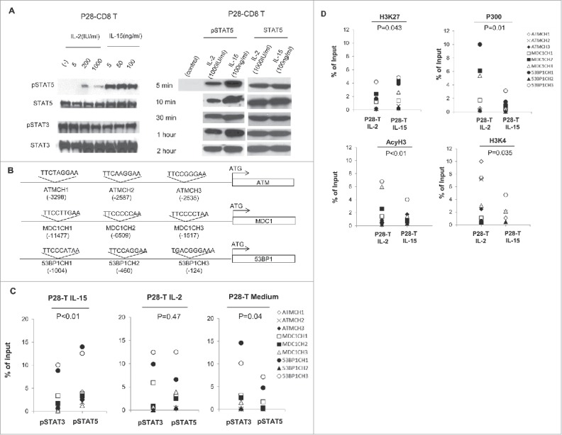
IL-15 strongly activates STAT5 signaling and changes the ratio of pSTAT5/3 signaling in CD8+ T cells. (A) Id-specific T cells starved of cytokines for 24 h were treated with IL-2 or IL-15 at different concentrations for 15 min. Total protein was extracted from the cytokine-treated T cells and equal amounts of protein were loaded into each lane. Anti-pSTAT3, anti-pSTAT5, anti-total STAT3, and anti-total STAT5 antibodies were used in Western blotting. (B) Schema for the nine potential STAT binding sites in the promoter regions of ATM, MDC1, and 53BP1. The prediction was carried out with TFSEARCH online program, and the potential STAT binding sequences and relative locations are indicated. (C) ChIP-PCR analysis of pSTAT5 and pSTAT3 binding to the STAT sites on the promoters of ATM, MDC1, and 53BP1 genes in cytokine-stimulated, day 14 IL-2- or IL-15-expanded Id L-chain-specific (P28) T cells, or unstimulated idiptype-specific CD8+ T cells. Shown are pooled data for nine STAT binding sites on the promoters of DNA damage genes. Isotype-matched antibodies were used as negative controls for all experiments (data not shown). (D) ChIP-PCR analysis of histones binding to the STAT sites on the promoters of ATM, MDC1, 53BP1 genes in day 14, IL-2- or IL-15-expanded idiotype L-chain specific T cells. H3K27: tri-methy-H3 (Lys27); P300: Histone acetyltransferase p300; H3K4: Histone tri methyl lysine 4; AcyH3: acetyl- Histone H3. MFI: Mean fluorescence intensity. p < 0.05.