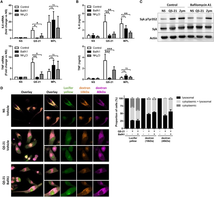
Figure 5.
QS-21-mediated dendritic cell activation and Syk phosphorylation depend on lysosomal maturation. (A,B) moDCs were treated with bafilomycin A1 (BafA1—250 nM) or NH4Cl (10 mM) for 1 h and stimulated with QS-21 (10 µg/ml) or MPL (1 µg/ml) for 4 h (A) or 24 h (B). TNF and IL-6 mRNA (A) and protein (B) were quantified by qPCR (n = 8) and ELISA (n = 11), respectively. Statistical significance was determined by two-way ANOVA followed by Tukey’s multiple comparisons test. (C) moDCs were treated with BafA1 for 1 h and stimulated with QS-21 (10 µg/ml) or zymosan as a positive control (Zym—50 µg/ml) for 2 h. Phospho-Syk (Y352), Syk, and β-actin were detected by sequential western blotting. One representative donor of 2 is shown. (D) moDCs were incubated with Lucifer Yellow, 10 kDa dextran-Alexa633, and 40 kDa dextran-Texas Red for 16 h then pretreated with BafA1 or DMSO (vehicle) and stimulated with QS-21 for 4 h. The localization of fluorescent markers was observed by confocal microscopy. The proportion of cells with punctate (lysosomal), diffuse (cytoplasmic), or both (punctate + diffuse) signals for the different fluorescent markers was determined with ImageJ. The data are representative of two donors.

