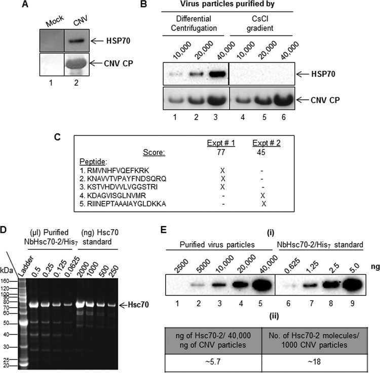
FIG 1.
Hsc70-2 is present in CNV virion preparations. (A) Western blot analysis of CNV virion preparations. Virion extraction was from equal masses of mock (lane 1)- and CNV (lane 2)-inoculated leaves. Pellets were resuspended in an equal volume of 10 mM sodium acetate (pH 5.8), and equal volumes of material (40,000 ng of CNV) were loaded onto a denaturing NuPAGE gel, followed by blotting to PVDF membranes. The upper panel was probed with an antibody that reacts to both Hsp70 and Hsc70 (here referred to as HSP70 antibody), and the lower panel was stained with Ponceau S to visualize CNV CP. (B) CNV virions (10,000, 20,000 and 40,000 ng) extracted by either differential centrifugation (lanes 1 to 3) or CsCl gradient centrifugation (lanes 4 to 6) were subjected to SDS-PAGE followed by Western blotting analyses. The upper panel shows a Western blot probed with an HSP70 antibody, and the lower panel shows the blot stained with Ponceau S to visualize CNV CP. (C) Summary of Hsc70-2 peptides detected by mass spectrometric analysis of proteins present in two independently purified CNV virion preparations (experiments [Expt] 1 and 2). The score shown for each experiment is based on a MASCOT search. X indicates the presence of the peptide in the mass spectrometric analysis. A BLAST analysis of the 5 detected peptides in the taxid Nicotianoideae identified 3 proteins that showed 100% identity to all five peptides (accession numbers AAP04522, AAR17080 and XP_009620324.1). All three proteins were most similar to Hsc70-2-like proteins as determined by BLAST analysis. The nucleotide sequences of these genes are most similar (95 to 96%) to that of N. benthamiana Hsc70-2 (Nbv5tr6412958; University of Sydney, Australia [http://benthgenome.qut.edu.au/]). These peptides were not detected in mock virus preparations extracted in a similar manner from the same amount of leaf tissue used to extract virions. (D) Denaturing gel electrophoresis of several dilutions (shown as microliters) of recombinant N. benthamiana Hsc70-2/His7 protein purified from bacterial cells using a Talon metal affinity matrix (Clontech). Bovine Hsc70 was used as the mass and size standard (shown in nanograms). The first lane shows a protein molecular mass standard (ladder) in kilodaltons. (E) Estimate of amounts of Hsc70-2 present in CNV virion preparations. Panel I, Western blot of CNV virion preparations probed with an HSP70 antibody. CNV particles (2,500, 5000, 10,000, 20,000, and 40,000 ng, as indicated) were electrophoresed under denaturing conditions and blotted to PVDF membranes prior to incubating with antibody. Dilutions of bacterially expressed N. benthamiana Hsc70-2/His7 (0.625, 1.25, 2.5, and 5.0 ng) probed with HSP70 antibody were used to estimate the amounts of Hsc70-2 present in CNV virion preparations. Panel ii, summary of amounts of Hsc70-2 present in virion preparations as determined by densitometry using Image Lab software version 5.1 (Bio-Rad). In an independent experiment, we found approximately 3.5 ng of Hsc70-2/His7 per 40 μg of virus (i.e., approximately 11 molecules of Hsc70-2 in 1,000 CNV particles).

