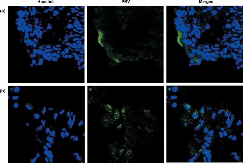
FIG 3.
PRV immunofluorescence staining. Immunofluorescence images of the nasal mucosa (a) and olfactory bulb (b) of the NIA3-inoculated 2-week-old piglet N08 euthanized at 3 days p.i. PRV antigens (green) were stained with a combination of mouse monoclonal anti-gB and -gD antibodies (1:100 in PBS) followed by an incubation step with FITC-labeled goat anti-mouse antibodies (1:200). Nuclei (blue) were counterstained with Hoechst 33342. Images were taken with a 63× objective on a confocal microscope.

