FIG 1.
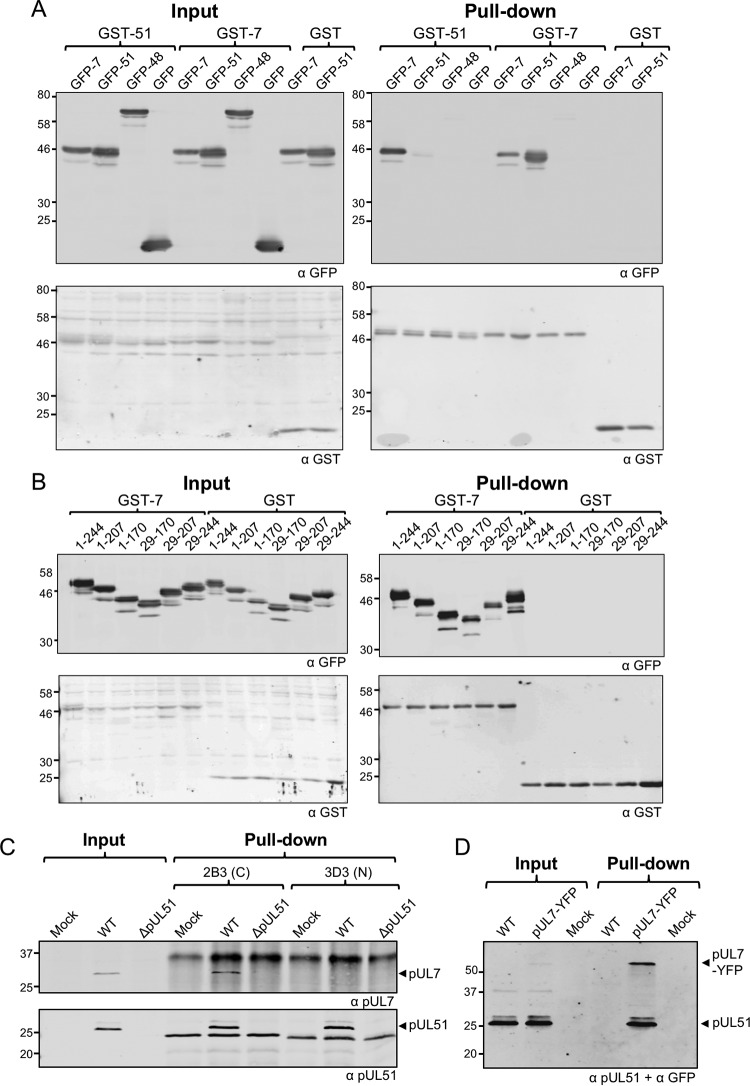
Interaction between pUL7 and pUL51 in transfected and infected cells. (A and B) 293T cells were cotransfected with plasmids carrying genes for GST-pUL51, GST-pUL7, or GST alone and GFP-pUL7, GFP-pUL51, GFP-pUL48, or GFP alone (A) or cotransfected with plasmids carrying genes for GST-pUL7 or GST alone, together with plasmids carrying genes for GFP-tagged full-length or truncated pUL51 (B). After 48 h, cells were lysed, and protein complexes were pulled down with glutathione-Sepharose beads. Proteins were separated by SDS-PAGE and immunoblotted, probing for GFP (top) and GST (bottom). (C) HaCaT cells were infected with WT or strain ΔpUL51 viruses and incubated for 16 h. Cell lysates were incubated with antibodies against pUL51, and the protein complexes were captured using protein A/G beads. The 3D3 antibody recognizes an epitope within the first 170 residues of pUL51, whereas 2B3 recognizes an epitope within residues 171 to 220 of pUL51. Samples were separated by SDS-PAGE and immunoblotted, probing for pUL7 (top) and pUL51 (bottom). (D) HaCaT cells were infected with WT or YFP-tagged pUL7 viruses and incubated for 16 h. Cell lysates were incubated with GFP-Trap–agarose beads to capture YFP-pUL7. Samples were separated by SDS-PAGE and immunoblotted, probing for pUL51 and YFP.

