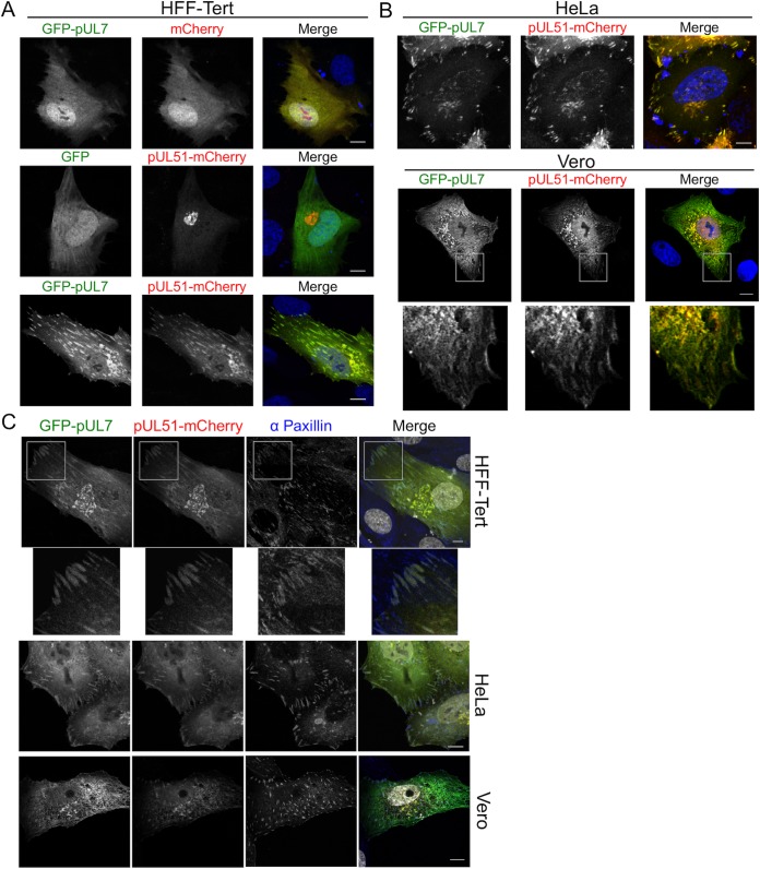
FIG 4.
pUL7 and pUL51 localization in transfected cells. (A and B) HFF-Tert cells (A) and HeLa or Vero cells (B) were cotransfected with GFP-pUL7 plasmid or empty GFP plasmid control and pUL51-mCherry plasmid or empty mCherry plasmid control in various combinations. After 24 h, cells were fixed and imaged by confocal microscopy. (C) HFF-Tert, HeLa, or Vero cells were cotransfected with GFP-pUL7 and pUL51-mCherry plasmids and fixed after 24 h. Cells were then immunostained for paxillin and imaged by confocal microscopy. Nuclei were stained with DAPI (blue in panels A and B and grayscale in panel C). Bars, 10 μm.

