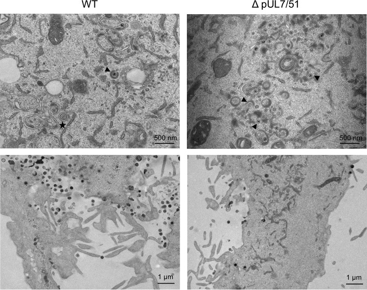
FIG 7.
Electron micrographs of WT and strain ΔpUL7-51 virus-infected HFF-Tert cells. Cells were infected with viruses for 16 h and processed for transmission electron microscopy imaging. The top panels show assembly sites, and bottom panels show secreted viral particles. Arrowheads indicate representative nonenveloped or partially wrapped cytoplasmic nucleocapsids, and the star indicates an enveloped virion within a vesicle.

