FIG 8.
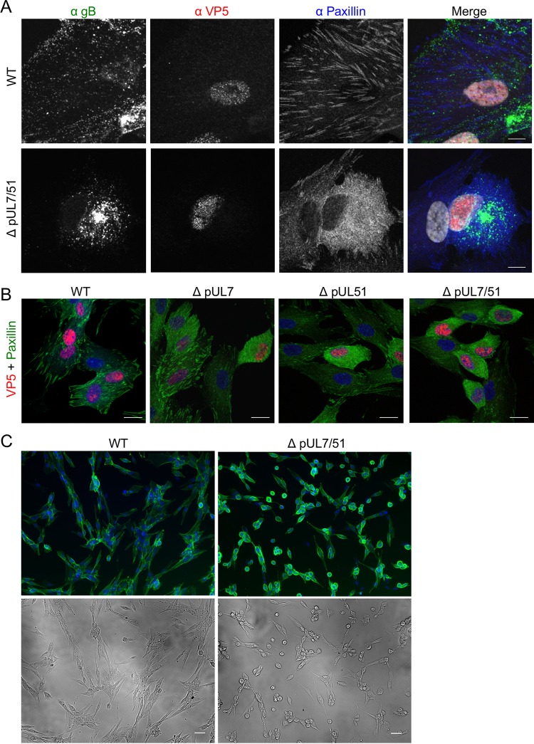
Protein localization and cell morphology in infected HFF-Tert cells. (A) Cells were infected with WT or ΔpUL7/51 strain virus and incubated for 8 h. After fixing, cells were labeled for gB, VP5, and paxillin. Bars, 10 μm. (B) Cells were infected with WT, ΔpUL7, ΔpUL51, or ΔpUL7/51 strain virus and incubated for 8 h. After fixing, cells were labeled for paxillin and VP5. Bars, 20 μm. (C) Cells were infected with either WT or ΔpUL7/51 strain viruses for 16 h, fixed, and then labeled for gD. Bars, 50 μm. Nuclei were stained with DAPI (grayscale in panel A and blue in panels B and C).

