FIG 1.

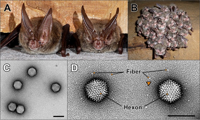
Host species from which bat adenovirus (BtAdV) 250-A was isolated and morphology of purified virions. (A) Two adult Rafinesque's big-eared bats (Corynorhinus rafinesquii) roosting in a building in Mammoth Cave National Park (MCNP), Kentucky, the site of isolation of BtAdV 250-A (photo courtesy of Tom Uhlman). (B) Rafinesque's big-eared bats hibernating in a cave in MCNP. Note the densely packed clusters formed by the bats during winter hibernation (photo courtesy of Steven Thomas, National Park Service). (C) Transmission electron micrograph of BtAdV 250-A virions negatively stained with uranyl formate, showing particles of icosahedral symmetry ∼90 nm in diameter (scale bar, 100 nm). (D) High-magnification view (52,476×) of BtAdV 250-A virions clearly showing the long flexible fiber proteins with their C-terminal knob domain visible (adjacent to the orange circles). Note an apparent kink (arrowhead) in the fiber protein, suggesting a potential hinge region imparting flexibility. Also highlighted is the hexon protein, which comprises the bulk of the adenovirus capsid, where 12 hexons form each of the 20 icosahedral facets of the particle (i.e., 240 copies per virion) (scale bar, 100 nm).
