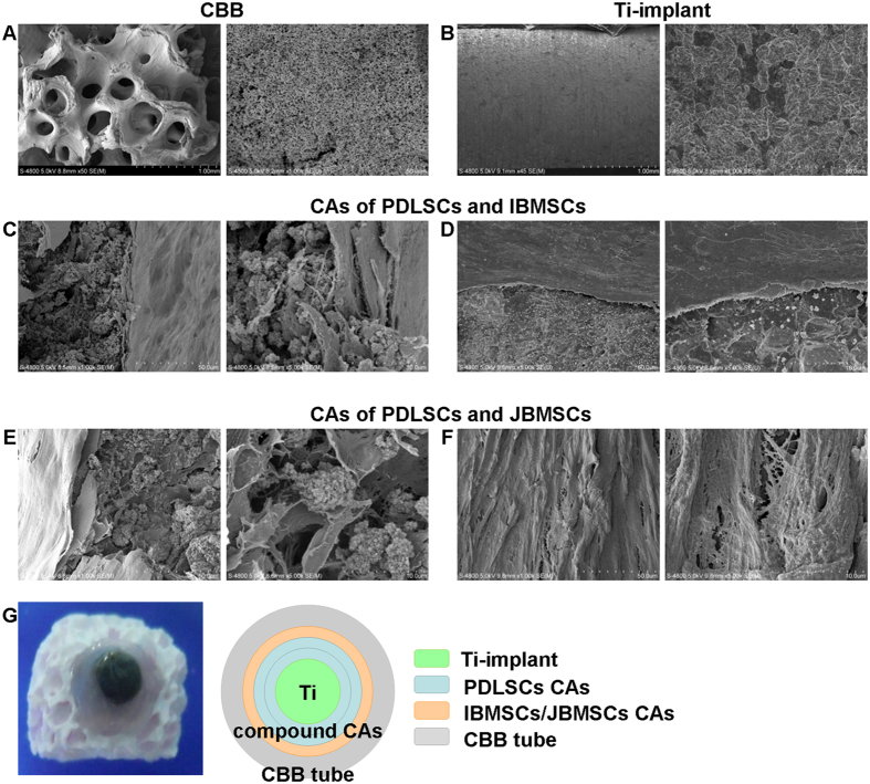
Figure 4. Microscopic appearance of the scaffold materials and the adhesion of the compound CAs to the scaffolds.
SEM images of CBB showed a porous structure, while Ti-implant showed a micro-pore facial structure (A,B). The attachment of compound CAs on the surface of CBB or Ti-implant was analyzed by SEM (C–F).

