
Figure 6.
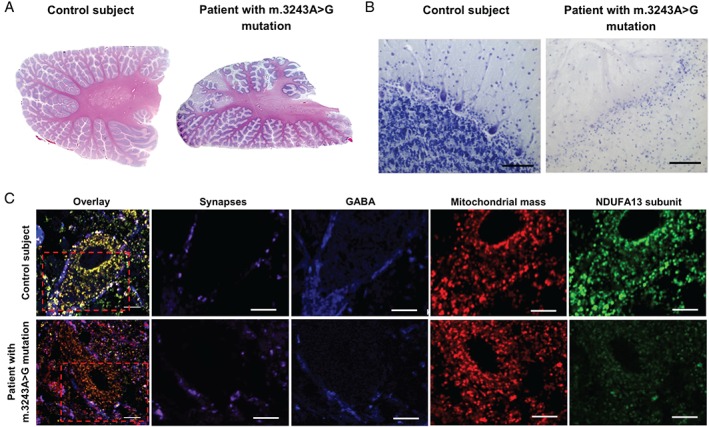
Cerebellar pathology in patients with the m.3243A>G mutation. (A) Numerous areas of necrosis are evident throughout the cerebellar cortex of a patient in comparison with control cerebellum (H&E staining). (B) Extreme neuronal loss is seen microscopically, affecting Purkinje cells and granule cells in the cortex (Cresyl fast violet staining). Scale bar: 100 µm). (C) In dentate nucleus neurons and in GABAergic (blue; GAD 65–67) synapses (magenta; synaptophysin) from Purkinje cells, there is downregulation of complex I (green; NDUFA13) relative to mitochondrial mass (red; COX4I2). Scale bar: 10 µm).