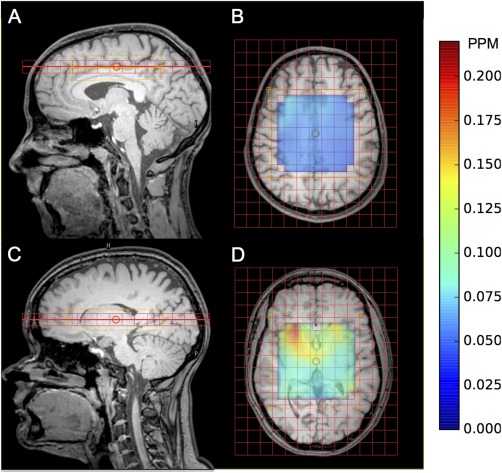
Figure 1.

Example healthy volunteer MRSI grid placement for: above the corpus callosum sagittal (A) and axial (B) views and the basal ganglia sagittal (C) and axial (D) views. Water line width map is included for corpus callosum (B) and basal ganglia (D) for the PRESS excitation region.
