
Figure 2.
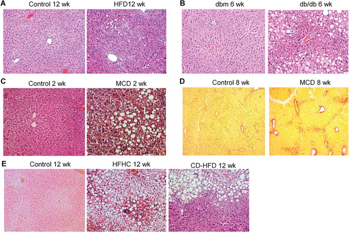
Histopathological features of NAFLD in different animal models. (A–C) Representative haematoxylin and eosin (H&E) staining of liver sections of: (A) C57BL/6 mice fed a control diet or an HFD for 12 weeks; (B) db/db and dbm control mice fed normal chow for 6 weeks; and (C) C57BL/6 mice fed a control diet or an MCD diet for 2 weeks. (D) Representative Sirius Red staining of liver sections of C57BL/6 mice fed a control diet or an MCD diet for 8 weeks. (E) Representative H&E staining of liver sections of C57BL/6 mice fed a control diet, an HFHC diet or a CD‐HFD for 12 weeks.