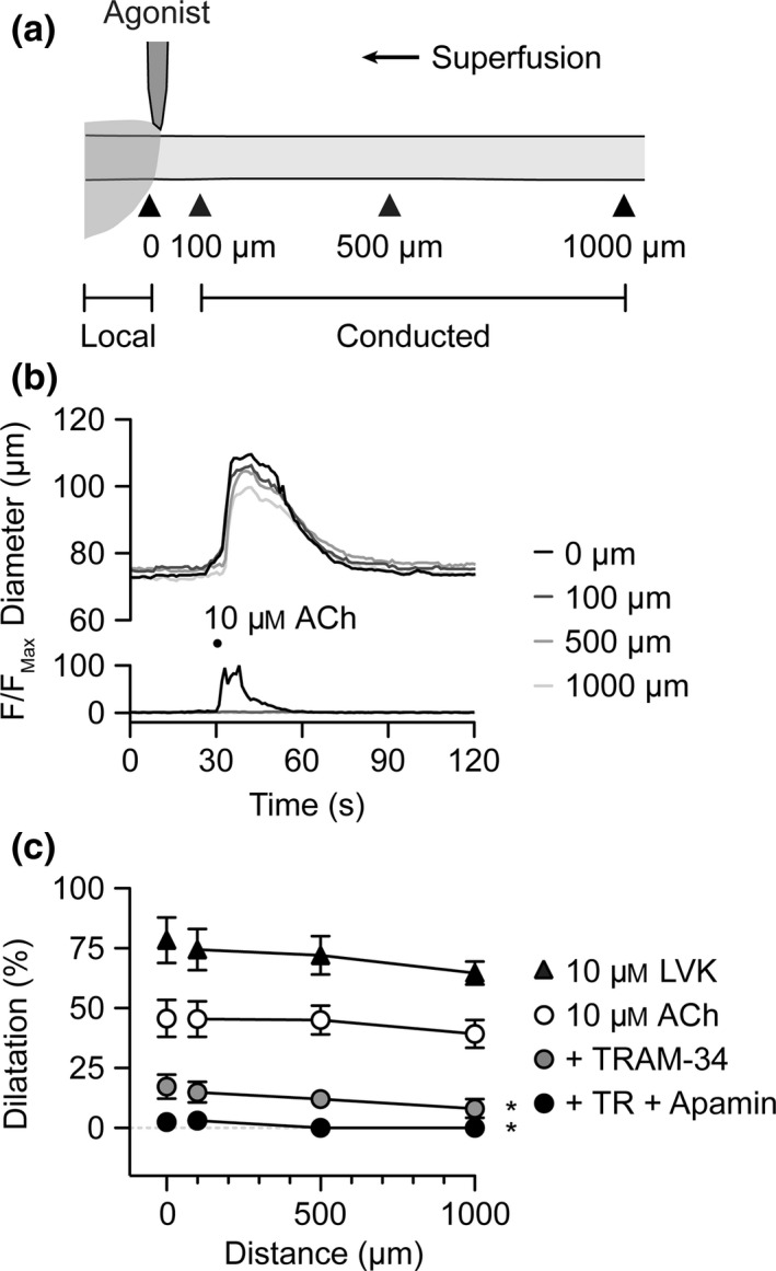
Figure 1.

IKC a channels underlie conducted dilatation to pulsed ACh. (a) Schematic of experimental set‐up for abluminal pressure‐pulse ejection of agonists to cannulated arterioles. The delivery micropipette was positioned at the downstream end of the arteriole. Diameter was measured locally (0 μm) and conducted responses at sites 100, 500 and 1000 μm upstream (indicated by arrowheads). (b) Representative time course of changes in diameter and fluorescence intensity in response to pulsed solution containing ACh (10 μ m) and carboxyfluorescein. (c) Summary data showing local and conducted responses to ACh under control conditions (white circles, n = 8), and during block of KCa channels by TRAM‐34 alone (n = 3) and with apamin (n = 3). In the same experiments, LVK (10 μ m) was subsequently pulsed to activate KATP channels (black triangles, n = 3). *P < 0.05 vs. 10 μ m ACh control. See Movie S1 for representative response to ACh.
