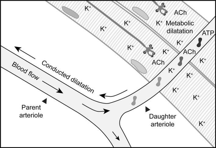
Figure 9.

Metabolic dilatation leads to conducted dilatation to improve blood flow. Schematic depicting the release of K+, ACh and ATP in response to contraction of a group of skeletal muscle fibres. The extracellular [K+] is elevated following repolarization and hyperpolarization of the skeletal muscle fibres and motor neurones, plus the arteriolar smooth muscle and endothelial cells. The ACh released from motor neurones can diffuse to the endothelium, and ATP released from circulating cells and endothelial cells themselves can act at endothelial cell P2Y receptors. Together, these autocrine and paracrine factors can ultimately hyperpolarize the arteriolar smooth muscle cells to stimulate local ‘metabolic’ dilatation, which can spread upstream to distal sites. The conducted dilatation further reduces vascular resistance, enabling improved flow into the area of high metabolic demand. For illustrative purposes, the skeletal muscle fibres are orientated to avoid direct contact with the parent arteriole, similar to the paradigm used for studies of focal electrical stimulation of skeletal muscle bundles (Cohen & Sarelius 2002).
