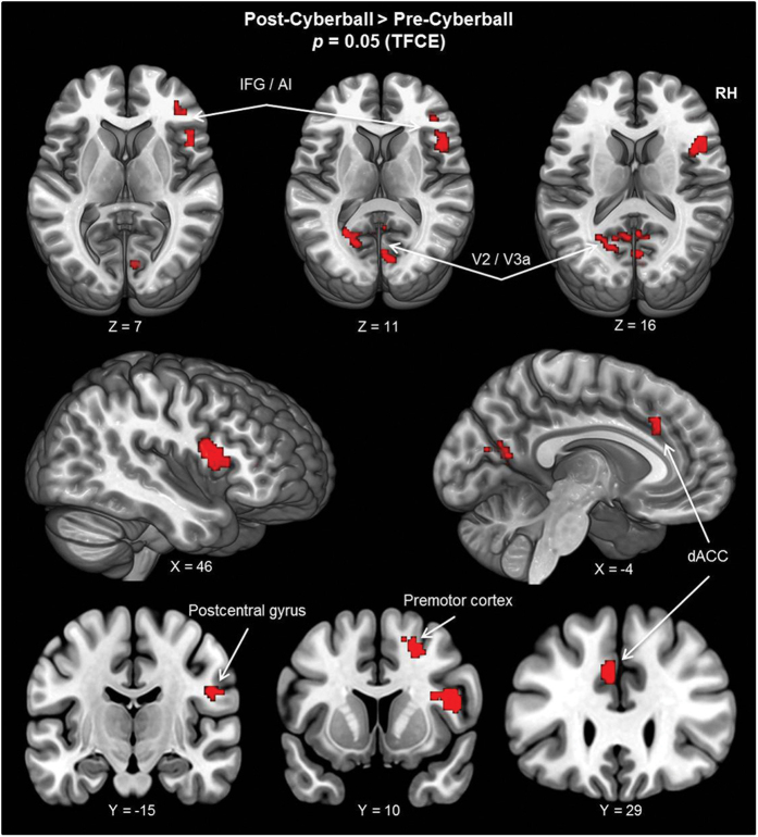
Figure 1. For the DMN, changes in resting-state functional connectivity between the two resting-state measurements are projected on the MNI template brain (ICBM 152).
Shown are all areas which exhibited increased functional connectivity with the DMN following the social stress-inducing cyberball task (i.e., post-cyberball > pre-cyberball). X, Y, and Z coordinates refer to MNI coordinates, indicating which slice is depicted. Thresholding and correction of multiple comparisons was achieved using the threshold-free cluster enhancement (TFCE) method, resulting in a whole-brain significant TFCE threshold of p < 0.05. (AI = anterior insula; dACC = dorsal anterior cingulate cortex; DMN = default mode network; RH = right hemisphere; IFG = inferior frontal gyrus).

