Figure 3.

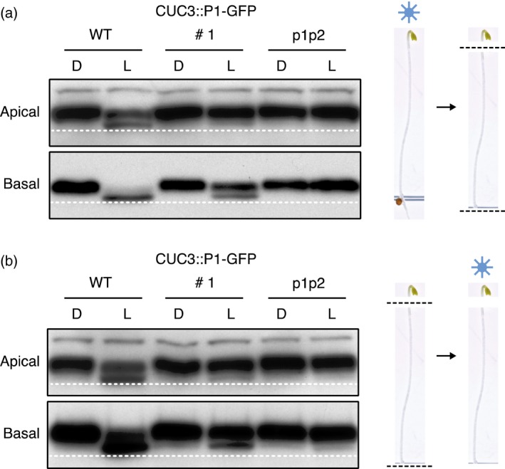
NPH3 phosphorylation status in apical and basal hypocotyl segments.
Immunoblot analysis of total protein extracts from 3‐day‐old etiolated wild‐type (WT) seedlings, seedlings expressing CUC3::PHOT1–GFP (CUC3::P1–GFP line 1) or the phot1 phot2 double mutant (p1p2). Seedlings were maintained in darkness (D) or irradiated with 20 μmol m−2 sec−1 of blue light for 15 min (L).
(a) Seedlings were dissected into apical and basal segments after blue light irradiation.
(b) Seedlings were dissected into apical and basal segments prior to blue light irradiation. Protein extracts were probed with anti‐NPH3 antibody. Dashed line indicates lowest mobility edge.
