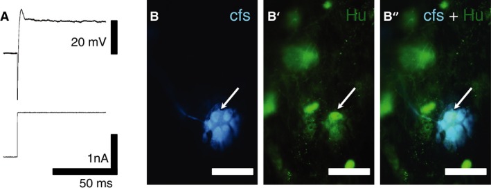
Figure 5.

Intracellular recording from a myenteric neuron with Hu‐translocation. (A) Depolarization with a large current pulse failed to evoke an action potential in this neuron. (B) Intracellular injection of carboxyfluorescein confirmed the neuronal morphology. The arrow indicates the nucleus. (B' and B”) Immunohistochemical labeling revealed translocation of Hu protein to the nucleus (arrows) (scale bar = 50 μm). Images were viewed on a Nikon confocal microscope (Eclipse Ti) with 20 × objective.
