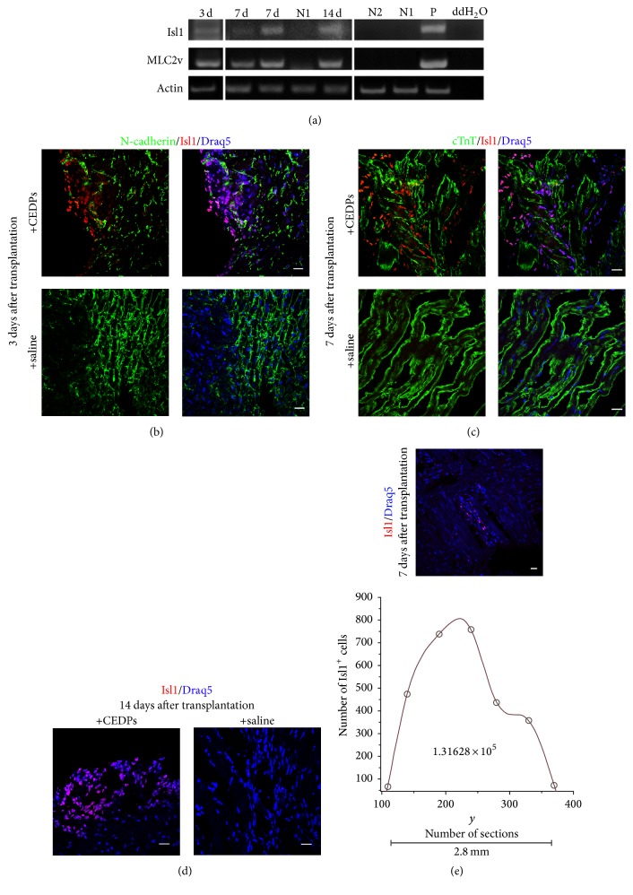
Figure 7.
Transplantation of CEDPs in adult rats' heart. (a) Expression of Isl1 and MLC2v after 3, 7, and 14 days of transplantation by RT-PCR analysis. mRNA from saline-injected immunosuppressed (N1) or untreated (N2) LV of adult rats was used as negative controls and mRNA from E12 mouse embryos (P) as positive control. (b–d) Clusters of Isl1+ cells derived from CEDPs were detected in frozen sections of LVs isolated 3, 7, and 14 days after transplantation. Sections were stained for DNA with Draq5 and with anti-N-cadherin (b) and anti-cTnT (c). Sections from isolated LVs of saline-injected immunosuppressed rats were used as control for each time-point. (e) All Isl1+ cells from two nonsequential sections were photographed (as in representative upper image) and counted with Fiji cell-counter. Draq5 used for DNA staining. The total Isl1+ cell number found in 2.8 mm tissue was calculated after spline interpolation. Scale bar: 20 μm.

