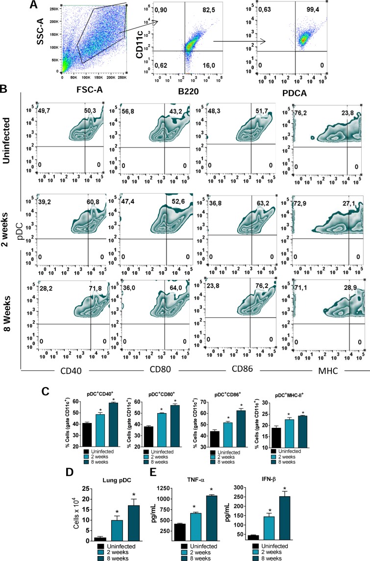
Fig 1. pDC response to P. brasilienis infection The influx of pDCs to the lungs of P. brasiliensis-infected mice (1×10⁶ yeasts cells) was determined by flow cytometry at weeks 2 and 8 post-infection.
Lungs were removed, leukocytes obtained and the number of pDCs evaluated. The pDCs were characterized as CD11c+B220+PDCA+ cells as indicated in the gate strategy used (A) and the activation measured by the expression of CD40, CD80, C86 and MHC-II molecules on their surface (B-C). The number of pDCs that migrated to the lungs was also determined by flow cytometric analysis (D). The levels of TNF-α and IFN-β were measured by ELISA in pDC supernatants obtained after18 hr of cell culture (E). Data represent the means ± SEM of at least 8 mice and are representative of two independent experiments with equivalent results (*p < 0.05).

