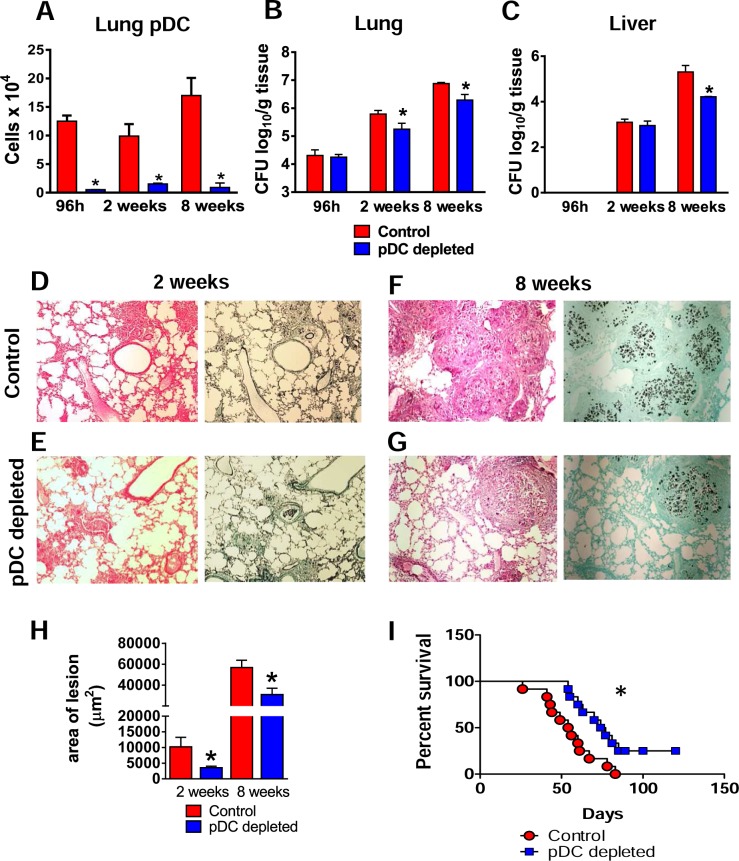
Fig 2. Depletion of pDCs reduces fungal loads, tissue injury and mortality rates.
Groups of anti-CD317 (anti-PDCA; clone BX44) or control rat IgG (clone HRPN) treated mice were infected i.t. with 1×10⁶ yeasts cells of P.brasiliensis. At 96 h, 2 and 8 weeks post-infection lungs were removed, leukocytes obtained and the numbers of pDC analyzed by flow cytometry (A). Colony-forming unit (CFU) counts from lungs (B) and liver (C) were determined 96 h, 2 and 8 weeks after P. brasiliensis infection. The bars represent means ± standard errors of the mean (SEM) of log10 CFU counts obtained from groups of 4–5 mice. (D–G) Photomicrographs of lung lesions of control (D and F) and pDC-depleted mice (E and G) at weeks 2 (D And E) and 8 (F and G) of infection. Lesions were stained with hematoxylin-eosin (left panels) and Grocott (right panels). (See also S1 Fig for liver lesions). (H) Total area of lesions in the lungs at week 2 and 8 of infection. (I) Survival curves of pDC-depleted and control infected mice were determined in a period of 110 days. Data represent the means ± SEM of at least 4 mice/group and are representative of two independent experiments with equivalent results (*p < 0.05).

