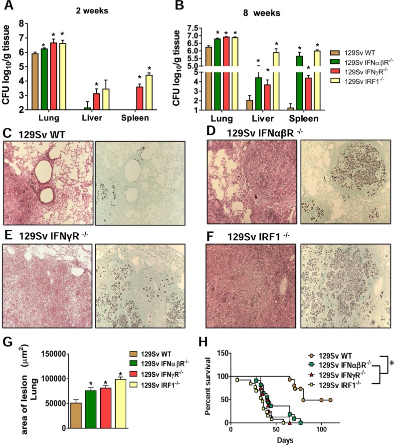
Fig 4. Absence of type I IFN signaling during P. brasiliensis infection increases mortality rates associated with increased fungal loads and tissue pathology.
Colony-forming unit (CFU) counts from organs were determined at 2 (A) and 8 weeks (B) after P. brasiliensis infection of 129Sv WT, IFNαβR−/−, IFNγR-/- and IRF1-/- mice. The bars represent means ± standard errors of the mean (SEM) of log10 CFU counts obtained from groups of 4–5 mice. Photomicrographs of lesions of WT mice (C), IFNαβR−/− (D), IFNγR-/- (E) and IRF1-/- (F) mice at week 8 of infection with 1 × 10⁶ P. brasiliensis yeasts. Staining of lesions was performed with hematoxylin-eosin (left panels) and Grocott (right panels). (G) Total area of pulmonary lesions at week 8 after infection. (H) Survival times of the four studied mouse strains infected i.t. with 1 × 10⁶ P. brasiliensis yeast cells were determined in a period of 110 days. Data represent the means ± SEM of at least 10 mice/group and are representative of two independent experiments with equivalent results. (*p < 0.05).

