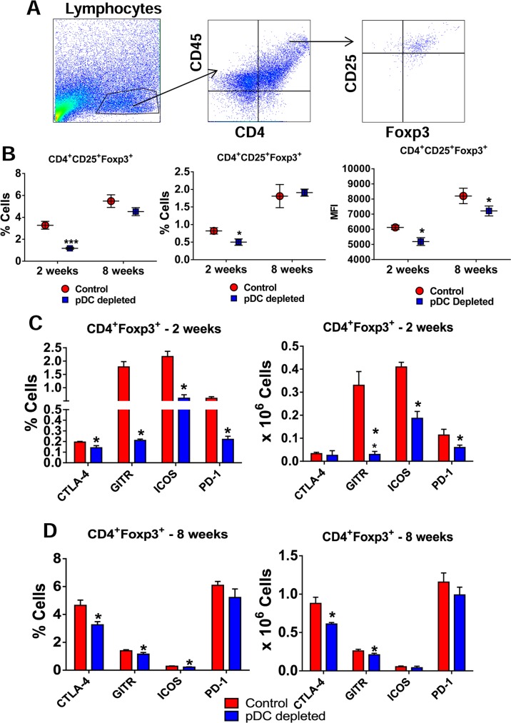
Fig 8. Depletion of pDCs impairs the influx of Treg cells to the lung of P. brasiliensis infected mice.
(A) Representative FACS plots demonstrating the gating strategy for CD4+ T and Treg cells. (B) Frequency (left panel), absolute numbers (central panel) and median fluorescence intensity (MFI, right panel) of CD4+Foxp3+ T cells analyzed by flow cytometry in the lungs of pDC-depleted and control mice after 2 and 8 weeks of infection. Bars reflect mean ± SD of three independent experiments with five mice per group (* p < 0.05). (C) Flow cytometric characterization of activation markers of CD4+Foxp3+ T cells such as CTLA-4, GITR, ICOS, and PD-1 expressed in frequency (left) and number (right) of cells in the lung infiltrating lymphocytes of pDC-depleted and control mice after 2 (C) and 8 weeks (D) of infection. Bars reflect mean ± SD of three independent experiments with five mice per group (* p < 0.05).

