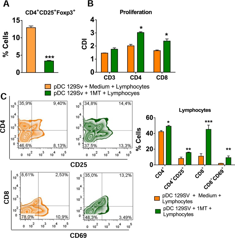
Fig 10. Impaired T cell responses promoted by pDCs is dependent on IDO activity.
pDCs were isolated from lungs of uninfected 129Sv mice using magnetics beads anti-mPDCA. The pDCs (5 x 10⁴) were treated or not with 1MT (1mM), matured with P. brasiliensis yeasts (1:10; Pb:pDC ratio) and then co-cultured for 7 days with splenic CD3+lymphocytes (1:10; pDC:lymphocytes ratio) isolated by anti-CD3 magnetic beads from WT mice. (A) Frequency of CD4+Foxp3+ T cells analyzed by flow cytometry. (B) Splenic lymphocytes from uninfected WT mice were previously labeled with CFSE (5 mM) and co-cultured with P. brasiliensis-infected pDCs. The co-culture was kept in the presence of RPMI medium containing or not 1MT. After 7 days, the cells were adjusted to 1 × 10⁶, labeled with specific anti-CD4 and CD8 antibodies and analyzed by flow cytometry. (C) After 7 days of co-culture with infected pDCs, lymphocytes were adjusted to 1 × 10⁶, labeled with specific anti-CD4, CD8, CD25, and CD69 antibodies and analyzed by flow cytometry. The lymphocytes were gated by FSC/SSC analysis and gated cells were analyzed for the expression of CD4+CD25+ (top) and CD8+CD69+ (bottom). Bars reflect mean ± SD of two independent experiments with eight mice per group (right) (* p < 0.05).

