Fig 3.
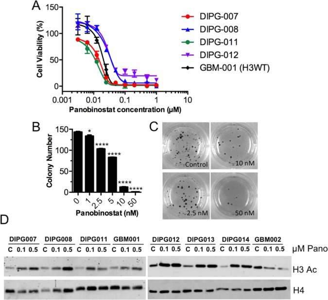
Panobinostat inhibits survival and clonogenicity of human DIPG cells in vitro (A) Human glioma cell models were treated with panobinostat for 72 h and then assayed for cell survival with an MTS Assay. Data are presented as the mean (with SD) of percent survival of control (untreated) cells. For each model, n = 6 replicates. (B-C) Human DIPG cells (HSJD-DIPG-007) were incubated with the indicated concentrations of panobinostat in soft agar for 2 weeks, and then colonies were stained with MTT and counted. Data in B are the mean with SEM, and statistical significance to compare drug concentration groups to controls were determined using an unpaired two-tailed t-test. * p = 0.02, **** p < 0.0001. Representative pictures of colonies are shown in C. (D) The indicated human glioma cell models were treated with either 0, 0.1, or 0.5 μM panobinostat for 48 h and then harvested for histone extraction. Histone lysates were separated via SDS-PAGE and blotted for the indicated antibodies.

